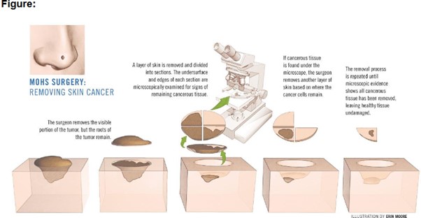
Mohs micrographic surgery (MMS) is a well-established specialised surgical method for removing skin cancer. It is primarily used for tumours on the head and neck. By using Mohs surgery, the surgical lesion is removed, mapped by colour-coded and thoroughly examined under the microscope. This treatment allows the entire peripheral and deep edges (margin) to be reviewed. If any tumour cells remain the process is repeated until the tissue is free of any tumour (see figure below). The procedure is done and repaired on the same day by the Mohs surgeon.
Diagram of removal of tumour tissue in a Mohs surgery
The benefits from Mohs Micrographic Surgery:
- Highest surgical success rate of 99% for skin cancers. Its effectiveness is due to the evaluation of the surgical margin and allowing the surgeon to trace/detect the extent of the “roots” of the cancer.
- Superior tissue conservation. This leads to optimising your repair to preserve natural function for an overall cosmetic result.
- The surgery is an outpatient day procedure. This ensures the patient that the cancer is entirely removed before they are sent home.
- Performed under Local anaesthesia which is fast acting with fewer risks rather than general anaesthesia.
The Day of Mohs Procedure
Mohs surgery are scheduled for an early start in the day. Patients should be aware that the surgery could potentially take the whole day. We suggest you may bring something to read or do to occupy your time in the recovery room. The patient can eat during the day of surgery, while waiting for the tissue analysis in our recovery area. We will supply lunch.
It is helpful to have someone to drive you home after your surgery. They may also accompany you for general support.
Upon your arrival you should check in at the reception desk. The procedure is done in our operating theatre. You will be escorted by our dermatology nurse who are able to answer any questions that you may have.
The Mohs surgeon will numb the area around the skin cancer using local anaesthetic. After this step, the remainder of the procedure is relatively pain free. The surgeon removes the layer for the Mohs technician to process. The removal of each layer takes one to two hours. Only about 20 minutes of that time is the actual surgical procedure. The remaining time being required for slide preparation and interpretation. Your wound will be dressed, and you will move to the recovery room whilst your tissue is getting processed.
The surgeon examines the entire surgical margin of the removed tissue under the microscope. In addition, Mohs surgery allows surgeons to treat poorly defined skin cancers or cancers that have been incompletely removed previously. On average, treated tumours require 2 stages for complete excision. If there is tumour found on the microscopic slides, the procedure is repeated until the tumour is removed.
Once the tissue is all clear of tumour, the surgeon will discuss reconstruction for your wound repair. The best method is determined on an individual basis after the defect is known. This will depend on the size and shape of the defect after the tumour is removed.